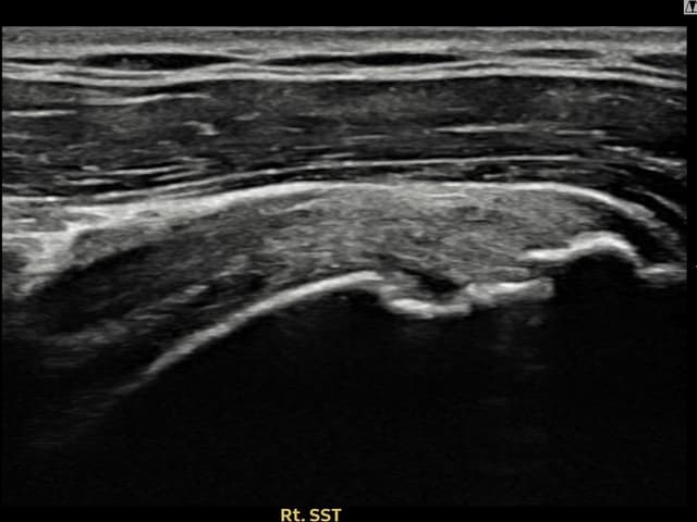
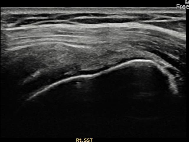
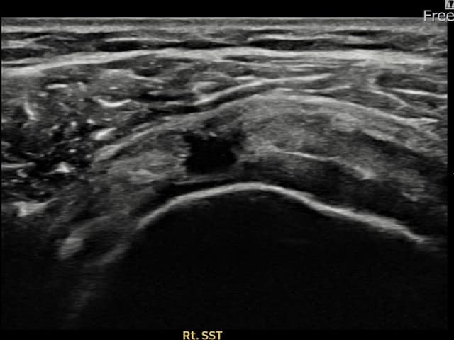
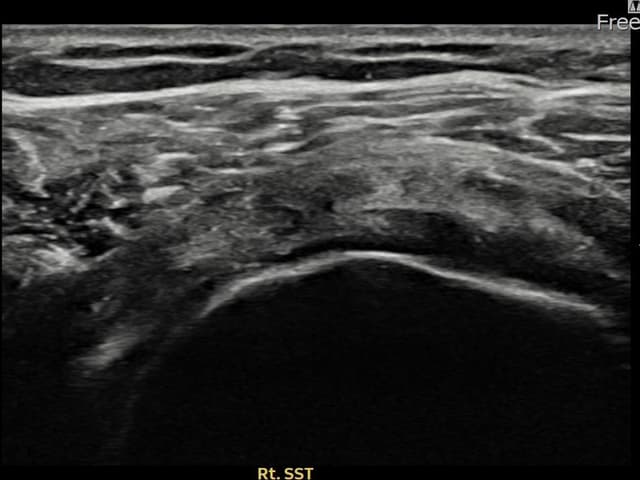
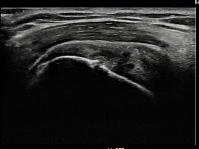
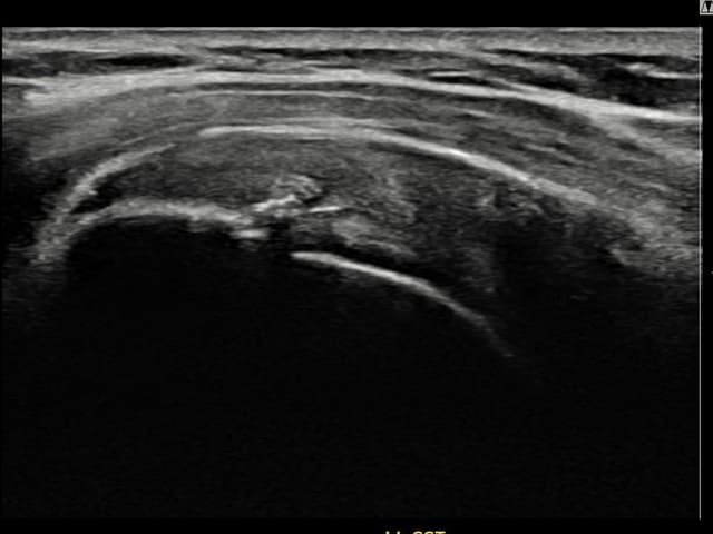
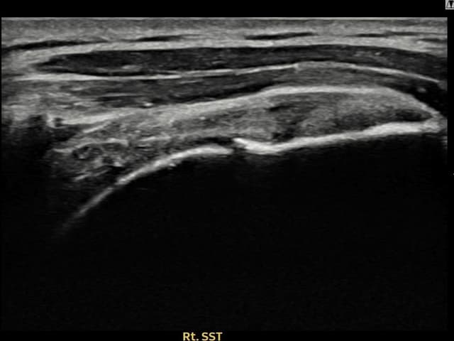
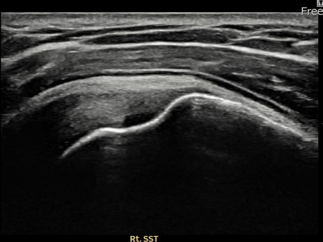
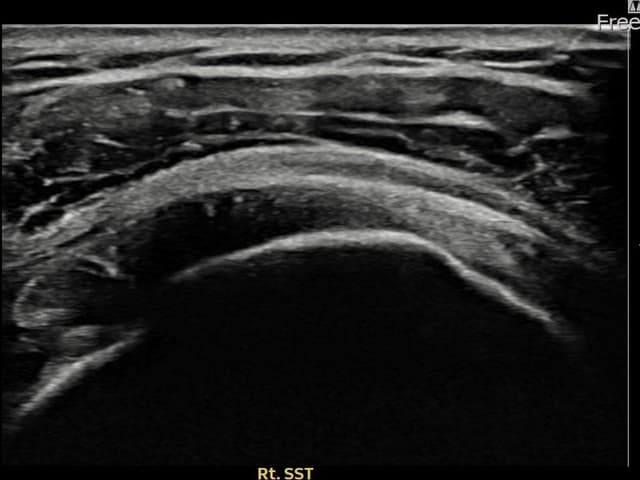
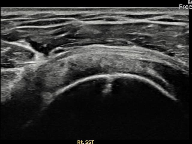
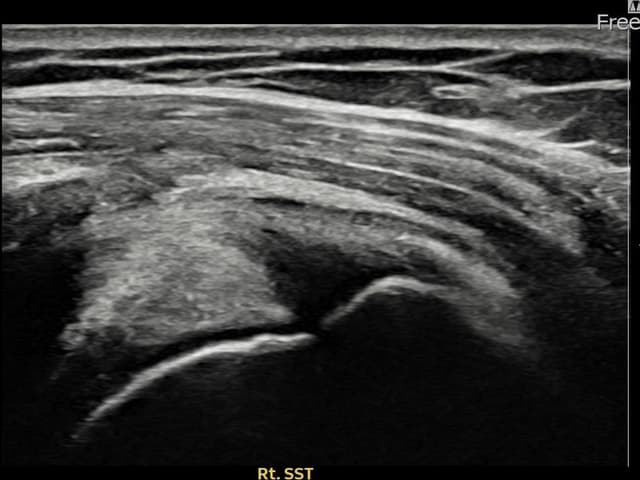
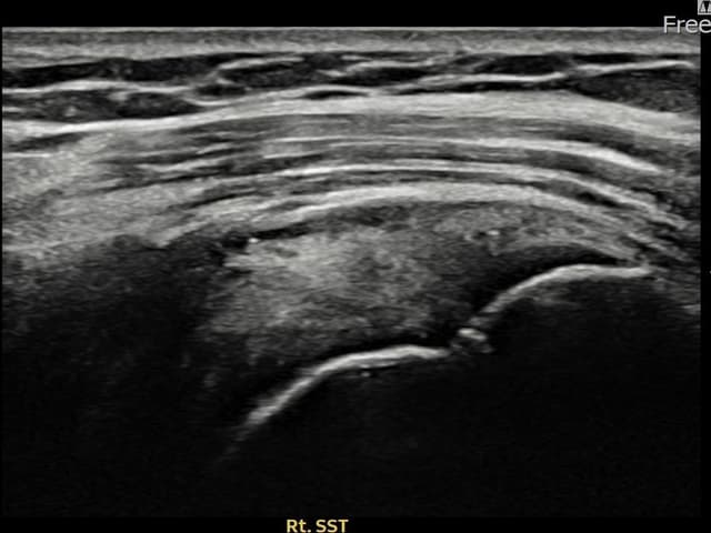
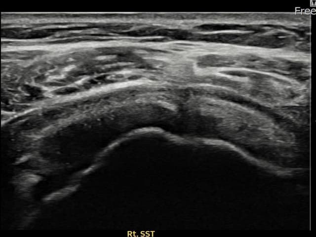
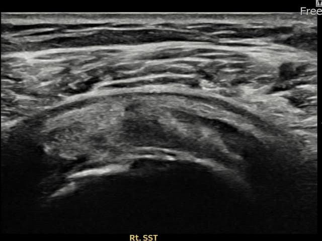
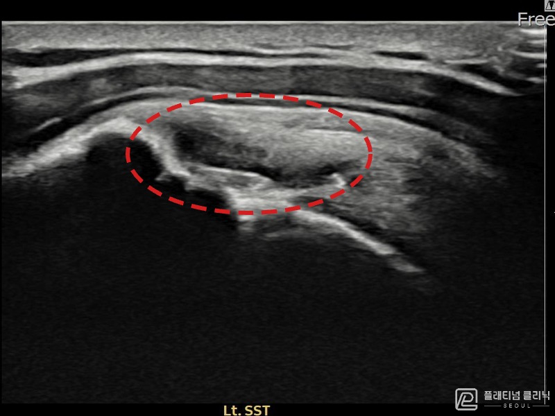
Treatment Results — Ultrasound Evidence
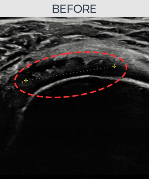
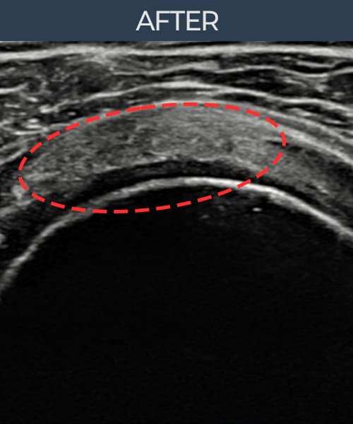
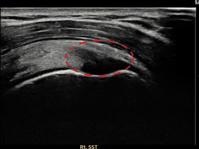
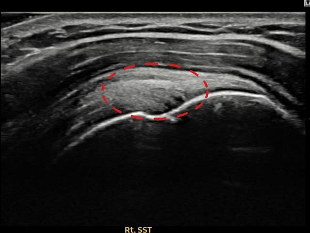
Real patient before-and-after ultrasound images.
See the recovery achieved without surgery.
All ultrasound images are published with patient consent. Individual results vary and identical outcomes are not guaranteed.
Total 30 cases
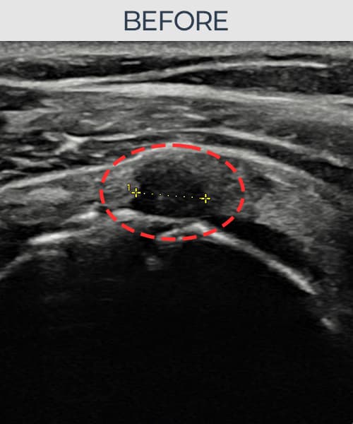
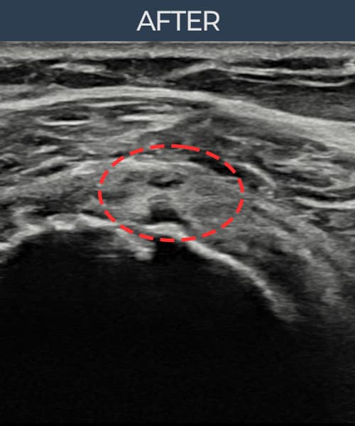
[Period: 25.10.14~26.01.08]
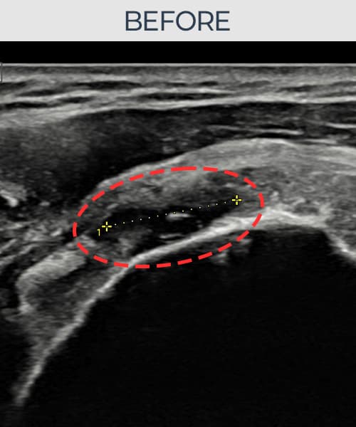
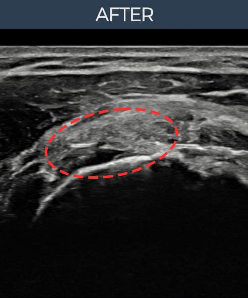
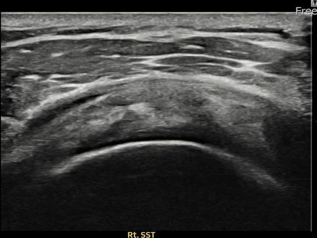
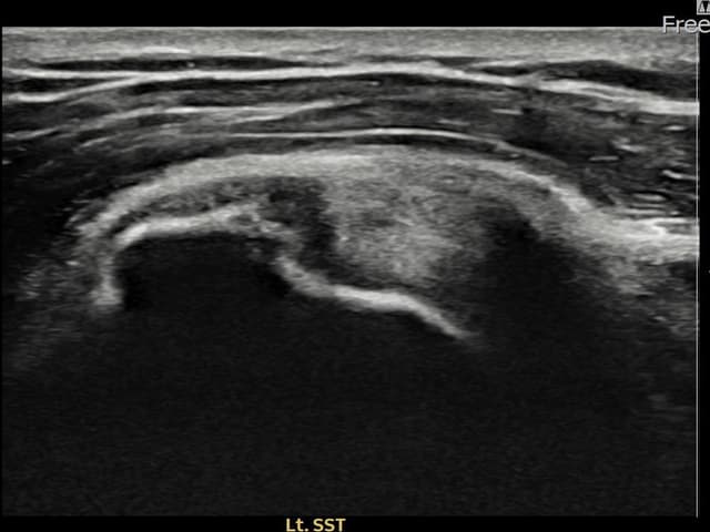
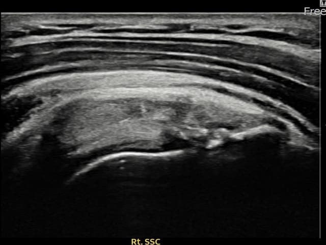
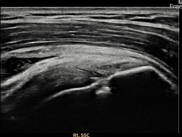
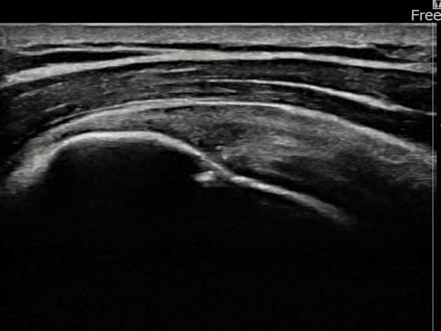
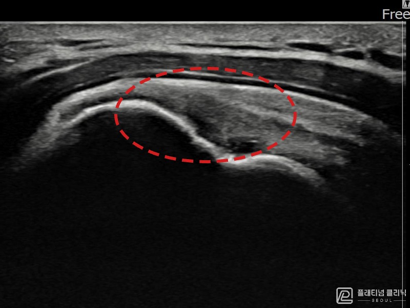
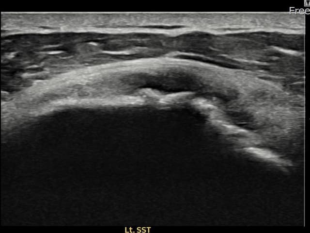
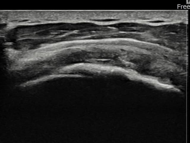
[Plication Suture] Ultrasound confirmed rotator cuff articular-side partial tear (14mm × 7mm (approx.
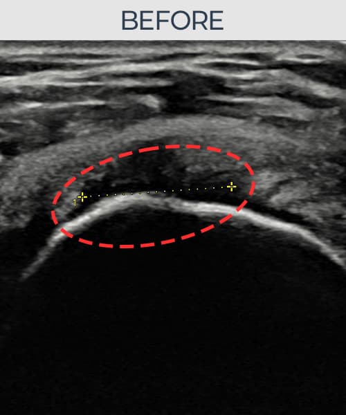
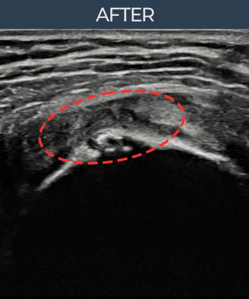
[Period: 25.07.02~25.09.24]
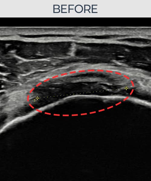
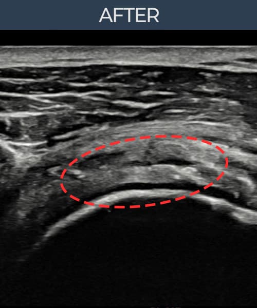
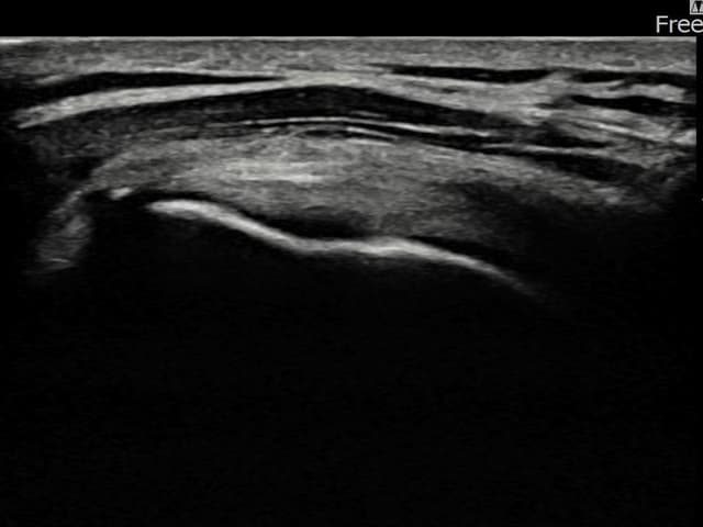
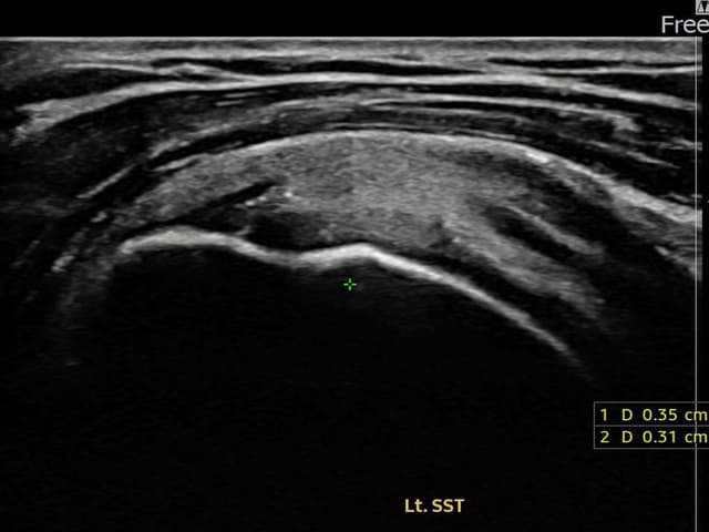
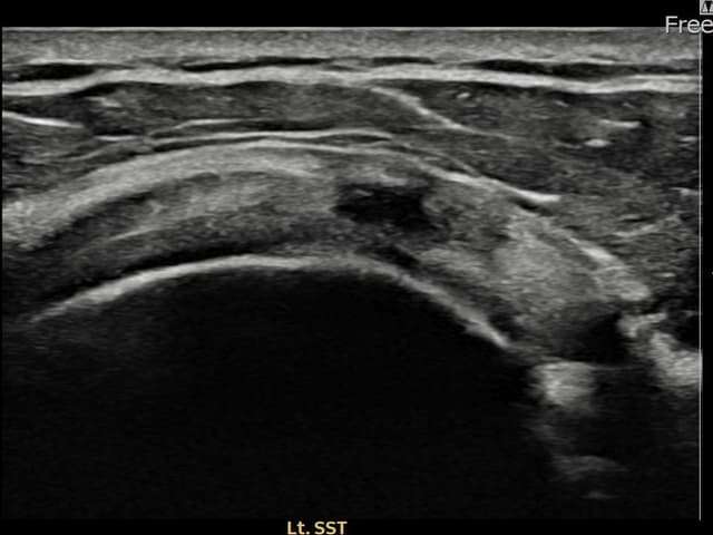
[Plication Suture] Ultrasound confirmed rotator cuff articular-side partial tear (8mm × 5mm (approx.
[Period: 25.03.10~25.06.04]
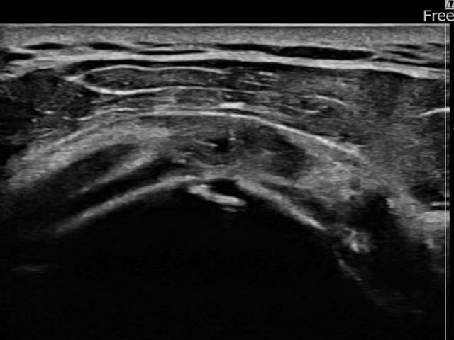
[Plication Suture] Ultrasound confirmed rotator cuff partial tear (6mm × 3mm (approx.
[Period: 24.11.04~25.01.14]
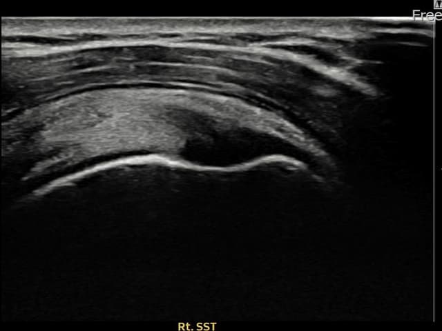
[Plication Suture] Ultrasound confirmed rotator cuff articular-side tear (9mm × 5mm (approx.
[Period: 24.10.14~24.12.23]
[Plication Suture] Ultrasound confirmed rotator cuff partial tear & ligament injury (11mm × 6mm (approx.
[Period: 24.09.23~24.12.03]
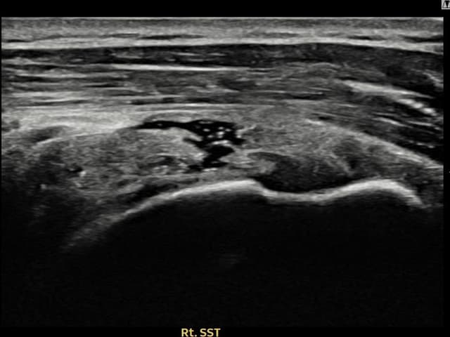
[Plication Suture] Ultrasound confirmed Right supraspinatus tendon articular-side partial tear (7mm × 4mm (approx.

[Period: 24.09.05~24.11.14]
[Plication Suture] Ultrasound confirmed Right supraspinatus tendon bursal-side partial tear (13mm × 6mm (approx.
[Period: 24.08.19~24.10.28]
[Plication Suture] Ultrasound confirmed Right supraspinatus tendon articular-side partial tear (10mm × 4mm (approx.
[Period: 24.07.29~24.10.07]
[Plication Suture] Ultrasound confirmed Right supraspinatus tendon articular-side partial tear (12mm × 4mm (approx.
[Period: 24.07.10~24.09.18]
[Plication Suture] Ultrasound confirmed Right supraspinatus tendon articular-side progressive tear (12mm × 7mm (approx.
[Period: 24.06.21~24.08.30]
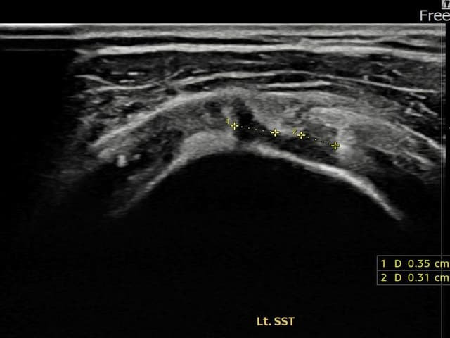
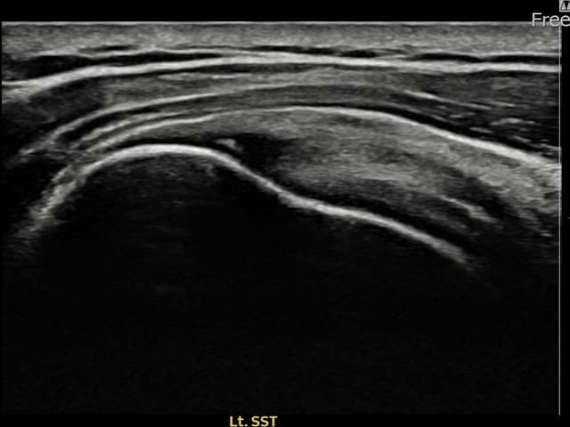
[Plication Suture] Ultrasound confirmed Left supraspinatus tendon articular-side partial tear (8mm × 3mm (approx.
[Period: 24.06.04~24.08.12]
[Plication Suture] Ultrasound confirmed Left supraspinatus tendon bursal-side partial tear (14mm × 7mm (approx.
[Period: 24.05.15~24.07.23]
[Plication Suture] Ultrasound confirmed Right supraspinatus tendon partial tear + 건내 석회화 (9mm × 4mm (approx.
[Period: 24.04.26~24.07.05]
[Plication Suture] Ultrasound confirmed Right supraspinatus tendon 관절면측 광범위 partial tear (16mm × 5mm (approx.
[Period: 24.04.09~24.06.17]
[Plication Suture] Ultrasound confirmed Left supraspinatus tendon footprint partial tear (11mm × 4mm (approx.
[Period: 24.03.21~24.05.29]
[Plication Suture] Ultrasound confirmed Right 견갑하근건 partial tear (8mm × 3mm (approx.
[Period: 24.03.04~24.05.10]
[Plication Suture] Ultrasound confirmed Right 견갑하근건 articular-side partial tear (7mm × 3mm (approx.
[Period: 24.02.14~24.04.22]
[Plication Suture] Ultrasound confirmed Left supraspinatus tendon partial tear (7mm × 3mm (approx.
[Period: 24.01.25~24.04.03]
[Plication Suture] Ultrasound confirmed Left supraspinatus tendon footprint partial tear (5mm × 3mm (approx.
[Period: 24.01.08~24.03.14]
[Plication Suture] Ultrasound confirmed Left supraspinatus tendon articular-side partial tear (3.
[Period: 23.12.14~24.02.22]
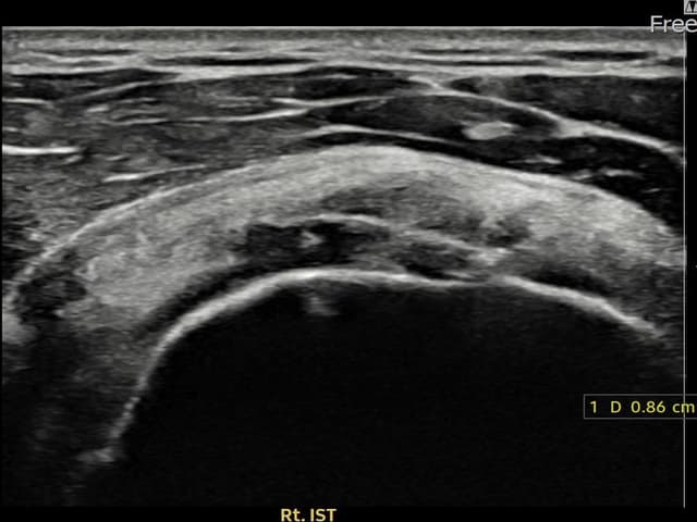
[Plication Suture] Ultrasound confirmed Right infraspinatus tendon footprint partial tear (8.
[Period: 23.11.28~24.02.06]
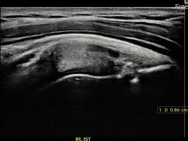
[Plication Suture] Ultrasound confirmed Right infraspinatus tendon articular-side partial tear (7mm × 3mm (approx.
[Period: 23.11.09~24.01.18]
[Plication Suture] Ultrasound confirmed Left supraspinatus tendon footprint partial tear (9mm × 4mm (approx.
[Period: 23.10.23~24.01.04]
[Plication Suture] Ultrasound confirmed Left supraspinatus tendon articular-side partial tear (8mm × 3mm (approx.
[Period: 23.10.05~23.12.07]
[Plication Suture] Ultrasound confirmed Right supraspinatus tendon footprint partial tear (10mm × 4mm (approx.
[Period: 23.09.19~23.11.21]
[Plication Suture] Ultrasound confirmed Right supraspinatus tendon articular-side partial tear (9mm × 4mm (approx.
[Period: 23.09.01~23.11.03]
[Plication Suture] Ultrasound confirmed Right supraspinatus tendon bursal-side partial tear (8mm × 4mm (approx.
[Period: 23.08.16~23.10.18]
[Plication Suture] Ultrasound confirmed Right supraspinatus tendon articular-side partial tear (7mm × 3mm (approx.
[Period: 23.07.31~23.09.27]
[Plication Suture] Ultrasound confirmed Left supraspinatus tendon 부착부 광범위 partial tear (13mm × 5mm (approx.
[Period: 23.07.18~23.09.14]
[Plication Suture] Ultrasound confirmed Left supraspinatus tendon 광범위 partial tear (15mm × 6mm (approx.
More cases available on Dr. Lee's blog and YouTube channel.
Book a Consultation
Dr. Lee personally assesses with ultrasound and plans your customized treatment