Rotator Cuff Plication Suture
Fix the Torn Tendon to Bone — Without Surgery
Especially effective for articular-side tearsthat don't improve with repeated injections. Shrinks the torn tendon and anchors it to the bone — no incision required.
What Is a Rotator Cuff Tear?
The rotator cuff consists of four tendons (supraspinatus, infraspinatus, teres minor, subscapularis) that wrap around the shoulder. A rotator cuff tear occurs when these tendons are damaged by wear or trauma.
Only part of the tendon thickness is torn. Classified as articular-side, bursal-side, or intrasubstance tear.
Non-surgical treatment possibleThe tendon is completely torn through. Categorized as small (1cm), medium (1–3cm), or large (3–5cm).
Judgment based on size and patternExtensive tear with retraction and fatty degeneration of the tendon.
Surgery may be requiredThe percentage alone doesn't tell the full story
“30% tear, 50% tear” — these numbers alone are not enough to determine the treatment direction. Which surface the tear is on and how it extends is the key to treatment selection. In particular, articular-side tears have structural reasons why injection therapy alone is unlikely to heal them.
Real Patient Case
“I've had injections for 6 months — why isn't it getting better? DNA injections, PDRN injections, collagen injections... Each time it seemed to get a little better, but the pain came back when I exercised. My doctor said 'It's an articular-side tear, so injections have their limits — consider surgery.' But surgery scared me...”
— Female patient in her late 30s, an avid badminton player
Why injections don't heal articular-side tears
It's like spraying adhesive on peeling wallpaper. No matter how good the ingredients, the wallpaper won't stay unless you press it back against the wall. Plication suture physically presses and anchors the tendon to the bone.
Why Injection Therapy Alone Doesn't Work
Poor Blood Supply
The articular side falls within the 'critical zone' — an area with sparse vascular distribution. Insufficient blood flow means inadequate nutrition, making natural healing extremely difficult.
Tendon-Bone Footprint Injury
This is where the tendon directly attaches to the bone (footprint). Once physically detached, simple injections cannot reattach it.
Progression to Full-Thickness Tear
Studies show that a significant proportion of articular-side partial tears progress to full-thickness tears over time. The risk is especially higher when the tear exceeds 50%.
Surgery vs Plication Suture
| Category | Arthroscopic Surgery | Plication Suture |
|---|---|---|
| Anesthesia | General anesthesia | Local (partial) anesthesia |
| Incision | 2–4 arthroscopic port incisions | None |
| Hospitalization | 1–3 days | Same-day discharge |
| Recovery | 3–6 months | 1–2 weeks (light daily activities) |
| Applicable tears | Full-thickness, massive tears | Partial tears, small full-thickness tears |
| Re-tear risk | Some exists | Tear progression suppressed |
| Cost | High (includes general anesthesia & hospitalization) | Relatively lower |
* Treatment approach may vary depending on tear type and patient condition. Optimal treatment is recommended after thorough evaluation.
Fundamentally Different from Standard Plication Suture
Bone micro-tunnel formation with a specialized drill
Standard plication suture anchors the suture only to soft tissue (the tendon itself). At Platinum, a specialized drill creates a micro-tunnel through the bone, allowing the suture to pass through and anchor to the bone. This provides far greater strength and stability than soft-tissue fixation alone.
Preventing tear progression is the key goal
One of the most important objectives of plication suture is to stop the tear from progressing. By suturing and stabilizing the current tear site, it prevents further damage. The earlier it is performed, the better the outcome.
Shrinkage and suture combined
The torn tendon is first shrunk (shrinkage) to reduce the size of the gap, then fixed to the bone with a suture. Both steps are performed together to fully stabilize the tear site.
Real-time ultrasound guidance
The entire procedure is performed under real-time ultrasound imaging. Dr. Lee confirms the exact position at every step, minimizing any risk to surrounding structures.
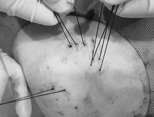
What Happens on the Day?
Precise tear evaluation and planning
MRI and ultrasound are used to analyze the tear location (articular-side vs bursal-side), tear direction, and footprint injury status. The suture path and bone tunnel position are planned in advance.
Local anesthesia (BPB)
Brachial plexus block anesthesia is administered to eliminate pain during the procedure. Consciousness is maintained — no general anesthesia required.
Bone micro-tunnel formation with specialized drill
Under real-time ultrasound guidance, a specialized drill creates a micro-tunnel through the bone. The suture is threaded through the tunnel to anchor the tendon to the bone.
Tendon shrinkage and suture fixation
The torn, widened tendon is shrunk (shrinkage) and secured to the bone tunnel with suture. Ultrasound confirms the tendon is closely approximated to the bone.
30 minutes of rest, then same-day discharge
After 30 minutes of rest post-procedure, the patient is discharged. An arm sling is worn for 2–4 weeks to protect the suture site, followed by a gradual rehabilitation program.
Ultrasound-Guided Plication Suture
Rotator cuff plication suture performed under real-time ultrasound guidance — no incision.
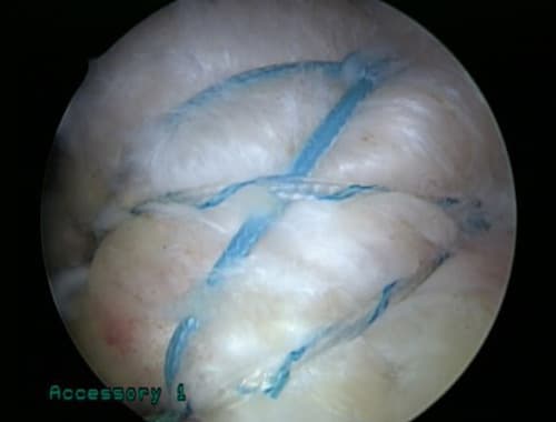
Suture Procedure
The process of shrinking and anchoring the torn tendon to the bone using specialized suture.
Suture Complete
The suture firmly anchors the tendon to the bone, stabilizing the tear site.
Multiplied Results with Combined Therapy
Plication suture is effective on its own, but combining it with regenerative therapy — depending on the tear type and condition — significantly enhances the speed and quality of tendon recovery after suture fixation.
After physically anchoring the tendon to the bone with suture, the patent-formula regeneration injection promotes collagen synthesis and tissue repair. Structural fixation + biological regeneration synergy.
After suture fixation, bone marrow stem cells are mobilized to the injury site to maximize tendon-bone junction regeneration. The suture provides solid support while stem cells do their work.
A bioabsorbable collagen patch is placed over the suture site to provide a regeneration scaffold. Tendon-bone integration stabilizes more rapidly.
About Suture Material Safety
The medical suture materials used in plication suture have decades of proven safety in surgical procedures worldwide.
Is it safe to leave the suture in my body?
Absorbable sutures are naturally absorbed within a few months. Non-absorbable materials are also biocompatible, safe for long-term coexistence with body tissue, and have been validated in hundreds of millions of surgical procedures worldwide.
Can an allergic reaction occur?
It is extremely rare but possible. A pre-procedure allergy history screening is conducted, and the material can be selected accordingly if needed.
Can I have an MRI after the suture?
This depends on the suture material used, but in general, MRI-compatible materials are selected. Specific guidance will be provided for any follow-up imaging after the procedure.
Plication Suture Q&A
I've had injection therapy for 6 months — why isn't it getting better?+
What happens if an articular-side tear is left untreated?+
Which type of tear responds best to plication suture?+
How is this different from surgery?+
Is the suture material safe? Is it okay to leave it in the body?+
Related Articles by Dr. Lee
Clinical Characteristics of Partial Rotator Cuff Tears
Reference
According to a systematic review published in Arthroscopy (2011), articular-side partial rotator cuff tears occur more than twice as frequently as bursal-side tears, and non-surgical treatment success rates reach 73–92% with appropriate patient selection. The study particularly emphasizes excellent functional recovery after non-surgical treatment for tears less than 50%.
Strauss EJ, et al. The Natural History of the Rotator Cuff. Arthroscopy. 2011;27(7):984-993. PMID 21482474 ↗Platinum Clinic's plication suture is a specialized non-surgical anchoring technique for articular-side partial tears, physically fixing the tendon to the bone through a micro-tunnel drilled in the bone. It is effective even when injection therapy has repeatedly failed.
Explanation Videos by Dr. Lee
Watch Dr. Lee Explain


Shoulder Tendon Tear — Check Before Surgery
If repeated injections haven't worked, plication suture may be the answer