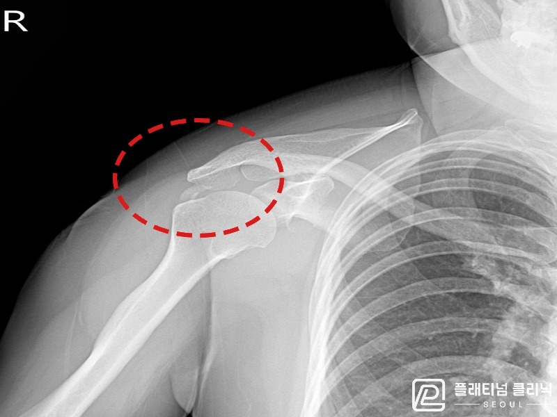
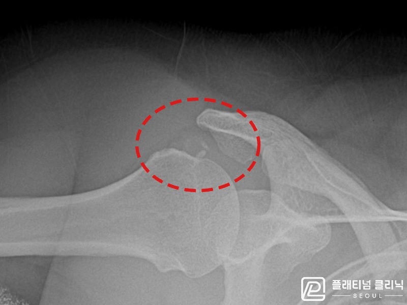
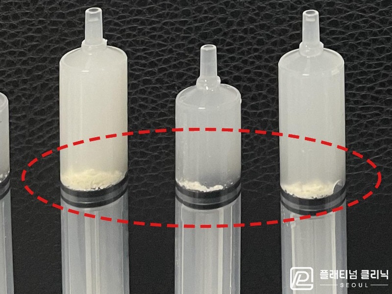
X-ray confirmed calcific deposits (11mm × 8mm) in the right supraspinatus tendon; ultrasound-guided calcific aspiration was performed. 6 syringes of calcium were aspirated, and follow-up X-ray at follow-up showed a marked reduction in calcific opacity.
[Period: 23.06.08]
Treatment Result
Pre-procedure X-ray confirmed calcium deposits (11mm × 8mm) in the right supraspinatus tendon; ultrasound also revealed a hyperechoic nodule with posterior acoustic shadowing. Under ultrasound guidance, calcific aspiration was performed — a total of 6 syringes of calcium were crushed and aspirated. Follow-up X-ray at follow-up showed a marked decrease in calcific opacity, with ongoing natural resorption of residual calcium confirmed.
Physician's Commentary
This patient presented with right shoulder pain due to calcific tendinitis confirmed on X-ray (deposit size: 11mm × 8mm). Given the size of the deposits and severity of symptoms, same-day ultrasound-guided calcific aspiration was performed. During the procedure, 6 syringes of calcium were successfully aspirated. Pain improved markedly from the procedure day or the following day. Follow-up imaging at follow-up confirmed significant reduction in calcific opacity, and the patient returned to daily activities without discomfort.
※ Published with prior patient consent. Photographed under identical conditions. Individual results may vary and side effects are possible — please consult your physician before proceeding with treatment.